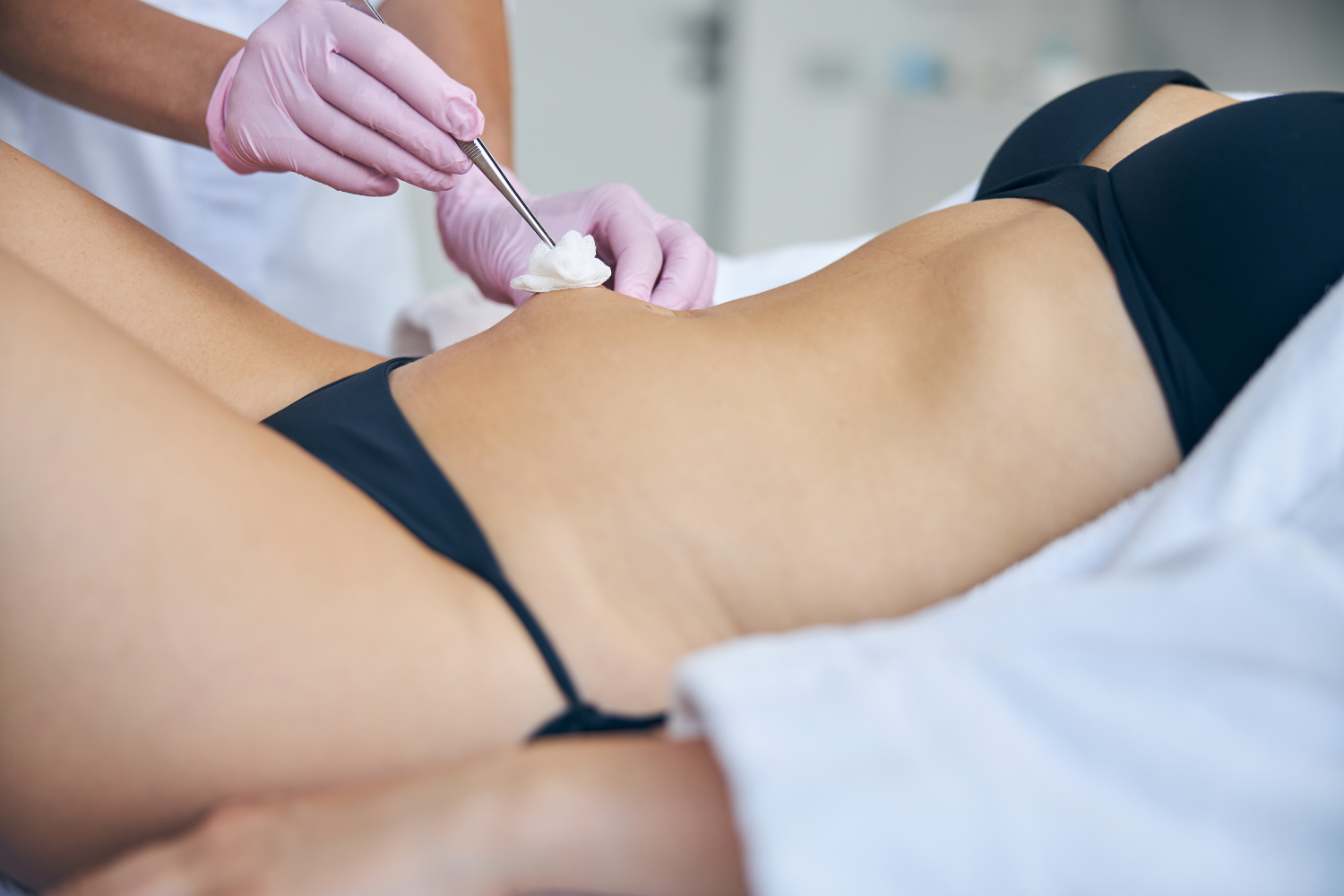
How safe is ultrasound guided fat transfer?

Ultrasound-guided fat transfer is an advanced technique that improves safety in aesthetic procedures, especially in the gluteal area. This technology allows the surgeon to see in real time where the fat is injected, avoiding errors that can cause serious complications. Thanks to this direct visualization, the risk of injecting fat into muscles or blood vessels decreases significantly.
By using ultrasound, the possibility of fat embolism, a complication that has been a major cause of mortality in these surgeries, is reduced. Recent studies show that the use of wireless ultrasound helps to keep the procedure within the subcutaneous space, improving both efficacy and safety for the patient. Therefore, this technique is gaining acceptance among professionals who seek to minimize risks.
Key points
- Real-time visualization improves accuracy and reduces risks.
- Avoid injecting into dangerous places such as muscles and blood vessels.
- Ultrasonic technology has demonstrated lower complication rates.
Importance of safety in ultrasound guided fat transfer
Safety is essential to avoid serious risks during fat transfer. Ultrasound allows the doctor to see precisely where the fat is being injected, helping to prevent deep tissue damage and complications.
Overview of fat transfer
Fat transfer involves removing fat from one part of the body and reinjecting it into another, for aesthetic or reconstructive purposes. This procedure may have risks if fat is injected into the wrong areas, such as large blood vessels.
The technique should prevent fat from entering the bloodstream, which could cause embolism. Therefore, the correct location of the injection is key to reducing these hazards. Accuracy improves results and protects patient health.
Advantages of using ultrasound in fat transfer
Real-time ultrasound allows you to visualize the layers of the skin and the tissues underneath. This helps the injection to be performed only in the subcutaneous area and not in deep planes, where important vessels are located.
With ultrasound, the doctor can adjust the needle and the amount of fat more precisely. Not only does this reduce risks, but it also improves fat distribution for a more natural result.
In addition, ultrasound makes it possible to detect possible complications during the procedure, making it possible to take immediate measures to ensure patient safety.
Safety Considerations Specific to Ultrasound
For a safe transfer with ultrasound, good planning and experience of the medical team is essential. Clear protocols should be used that include asepsis measures and informed consent.
The operator must have solid training in ultrasound techniques to interpret images correctly. It is also necessary to ensure that the devices are calibrated and that the procedure is stopped if a risk is detected.
Ultrasound does not eliminate all risks, but it reduces critical events. Post-procedure follow-up and evaluation are key to confirming that there are no late complications.
Protocols and Practices for Safe Fat Transfer
Fat transfer safety depends on a careful process from patient selection to the technique used. It is essential to prepare the patient, maintain a clean environment and avoid hazardous areas during the injection. These steps reduce risks and improve outcomes.
Patient Selection and Evaluation
The first step is to evaluate the patient's general health. You must be in good physical condition, without diseases that affect healing or coagulation. It's key to review medical history and allergies. The quantity and quality of the fat available for transplantation is also analyzed.
The patient must understand the real risks and expectations. It is recommended to avoid surgery for people with serious heart problems or uncontrolled diabetes. This selection reduces complications and ensures that the procedure is safe and effective.
Aseptic and preparation techniques
Before starting, the surgeon must prepare the area with rigorous cleansing to prevent infection. Antiseptic solutions are used to clean the extraction area and the receiving area. In addition, the equipment must be sterilized and ready for the procedure.
The use of real-time ultrasound allows the cannula to be guided to inject fat into the correct layer. Local or general anesthesia is administered on a case-by-case basis, with constant monitoring. Everything must be done while minimizing tissue trauma to facilitate good fat integration.
Identifying and avoiding risk structures
During the injection, it is vital to avoid deep blood vessels and muscles. Ultrasound technology helps to visualize these structures and ensures that fat is placed in the subcutaneous layer.
Placing fat in the correct plane reduces risks such as embolism or nerve damage. The surgeon must move slowly and control pressure so that the material is deposited evenly and not in vessels or muscles. This practice improves safety and post-operative recovery.
Potential complications and prevention
The most common complications of ultrasound-guided fat transplantation include seromas, infections, and the risk of fat embolism. Prevention is based on accurate technique and constant monitoring during and after the procedure.
Early recognition of complications
Detecting complications in their early stages is crucial. Warning signs are excessive swelling, unusual pain, or redness in the treated area.
The surgeon should perform frequent evaluations to identify seromas or infections. The seroma presents as an accumulation of fluid, detectable by ultrasound.
Key Indicators:
- Persistent swelling
- Localized pain
- Changes in skin color
Rapid identification allows for timely treatment and reduces major risks.
Managing Fat Infections and Embolisms
Infections occur at a low percentage, around 0.8%, and are treated with antibiotics adjusted according to severity. It is vital to maintain a sterile technique during the procedure.
Fat embolism, although rare, is the most serious complication. Using ultrasound to guide the injection in the subcutaneous plane prevents fat from entering deep veins or muscles, reducing this risk.
Key Practices for Preventing Fat Embolism:
- Avoid intramuscular injection
- Use real-time images
- Control the pressure and volume injected
Postoperative follow-up and continuous evaluation
After surgery, the patient must be monitored regularly. Visits make it possible to evaluate healing, detect complications early and adjust care.
It is important for the patient to report any unusual symptoms. Ultrasound may be used to check the area and confirm that there is no fluid accumulation or signs of infection.
Monitoring guidelines:
- Weekly review for the first month
- Clinical and imaging exams
- Patient education on warning signs
This follow-up reduces complications and ensures better long-term outcomes.
Frequently Asked Questions
Ultrasonic guidance in fat transplantation allows for precise tissue placement in the subcutaneous space. This method reduces significant risks and helps control absorption and complications associated with the procedure.
What is the safety level of the ultrasound guided fat transplant procedure?
The ultrasound-guided procedure improves safety by allowing for real-time visualization. This prevents the injection of fat into the muscle, reducing the risk of serious complications.
What complications can arise from an ultrasound guided fat transplant?
Although ultrasound minimizes risks, complications such as infection, skin irregularities, or irregular tissue absorption can occur. Early visualization helps detect and manage them.
Is fat tissue absorption common after an ultrasound guided transplant?
Yes, some of the tissue can be reabsorbed by the body over time. Ultrasonic guidance allows fat to be distributed more evenly, which can decrease the amount of absorption.
What are the risks associated with the transfer of fat to the breasts?
Risks include cyst formation, changes in breast texture and, in rare cases, vascular complications. The ultrasound technique helps to avoid injection into large blood vessels.
What precautions should be taken before undergoing an ultrasound-guided fat transfer?
A full medical evaluation and a discussion of a health history is recommended. The surgeon's experience in the technique and proper use of ultrasound are key to patient safety.
What ultrasound technique is used to ensure a safe and effective fat transplant?
A real-time image is used with a wireless device that allows us to visualize both tissues and blood vessels. This ensures that fat is deposited only in the subcutaneous space avoiding deep tissues.
Safety in surgery is non-negotiable, technique is everything
Ultrasound-guided fat transfer not only improves aesthetic results, but it marks a before and after in the safety of the procedure. By visualizing the patient's anatomy in real time, Dr. Antonio García Rodríguez can position fat with millimeter precision, always on safe planes and without jeopardizing vital structures.
This level of control is not a luxury, but rather a standard that reflects a commitment to your short and long term well-being.
The best decision is not just to look good, but to do so with high-level technical and surgical support.


